Brachial Plexus Injury: A Nerve Injury You Shouldn't Ignore.

The brachial plexus is the network of nerves that sends signals from your spine to your shoulder, arm and hand. A brachial plexus injury occurs when these nerves are stretched, compressed, or in the most serious cases, ripped apart or torn away from the spinal cord.

Minor brachial plexus injuries, known as stingers or burners, are common in contact sports, such as football. Babies sometimes sustain brachial plexus injuries during birth. Other conditions, such as inflammation or tumors, may affect the brachial plexus.

The most severe brachial plexus injuries usually result from auto or motorcycle accidents. Severe brachial plexus injuries can leave your arm paralyzed, with a loss of function and sensation. Surgical procedures such as nerve grafts, nerve transfers or muscle transfers can help restore function.
 

Symptoms

Signs and symptoms of a brachial plexus injury can vary greatly, depending on the severity and location of your injury. Usually only one arm is affected.

Less severe injuries

Minor damage often occurs during contact sports, such as football or wrestling, when the brachial plexus nerves get stretched or compressed. These are called stingers or burners, and can produce the following symptoms:

  • A feeling like an electric shock or a burning sensation shooting down your arm
  • Numbness and weakness in your arm

These symptoms usually last only a few seconds or minutes, but in some people may linger for days or longer.

More-severe injuries

More-severe symptoms result from injuries that seriously injure or even tear or rupture the nerves. The most serious brachial plexus injury (avulsion) occurs when the nerve root is torn from the spinal cord.

Signs and symptoms of more-severe injuries can include:

  • Weakness or inability to use certain muscles in your hand, arm or shoulder
  • Complete lack of movement and feeling in your arm, including your shoulder and hand
  • Severe pain

When to see a doctor

Brachial plexus injuries can cause permanent weakness or disability. Even if yours seems minor, you may need medical care. See your doctor if you have:

  • Recurrent burners and stingers
  • Weakness in your hand or arm
  • Weakness in any part of the arm following trauma
  • Complete paralysis of the upper extremity following trauma
  • Neck pain
  • Symptoms in both arms
  • Symptoms in upper and lower limbs

It's important to be evaluated and treated within six to seven months after the injury. Delays in treatment may compromise outcomes of nerve surgeries.

Causes

Damage to the upper nerves that make up the brachial plexus tends to occur when your shoulder is forced down while your neck stretches up and away from the injured shoulder. The lower nerves are more likely to be injured when your arm is forced above your head. These injuries can occur in several ways, including:

  • Contact sports. Many football players experience burners or stingers, which can occur when the nerves in the brachial plexus get stretched beyond their limit during collisions with other players.
  • Difficult births. Newborns can sustain brachial plexus injuries when there are problems during birth, such as a breech presentation or prolonged labor. If an infant's shoulders get wedged within the birth canal, there is an increased risk of a brachial plexus palsy. Most often, the upper nerves are injured, a condition called Erb's palsy. Total brachial plexus birth palsy occurs when both the upper and lower nerves are damaged.
  • Trauma. Several types of trauma — including motor vehicle accidents, motorcycle accidents, falls or bullet wounds — can result in brachial plexus injuries.
  • Inflammation. Inflammation may cause damage to the brachial plexus. A rare condition known as Parsonage-Turner syndrome (brachial plexitis) causes brachial plexus inflammation with no trauma and results in paralysis of some muscles of the arm.
  • Tumors. Noncancerous (benign) or cancerous tumors can grow in the brachial plexus or put pressure on the brachial plexus or spread to the nerves, causing damage to the brachial plexus.
  • Radiation treatment. Radiation treatment may cause damage to the brachial plexus.

Risk factors

Participating in contact sports, particularly football and wrestling, or being involved in high-speed accidents increases your risk of brachial plexus injury.

Complications

Given enough time, many brachial plexus injuries in both children and adults heal with no lasting damage. But some injuries can cause temporary or permanent problems:

  • Stiff joints. If you experience paralysis of your hand or arm, your joints can stiffen, making movement difficult, even if you regain use of your limb. For that reason, your doctor is likely to recommend ongoing physical therapy during your recovery.
  • Pain. This results from nerve damage and may become chronic.
  • Loss of feeling. If you lose feeling in your arm or hand, you run the risk of burning or injuring yourself without knowing it.
  • Muscle atrophy. Slow-growing nerves can take several years to heal after injury. During that time, lack of use may cause the affected muscles to break down (degenerate).
  • Permanent disability. How well you recover from a serious brachial plexus injury depends on a number of factors, including your age and the type, location and severity of the injury. Even with surgery, some people experience permanent disability, ranging from weakness in the hand, shoulder or arm to paralysis.

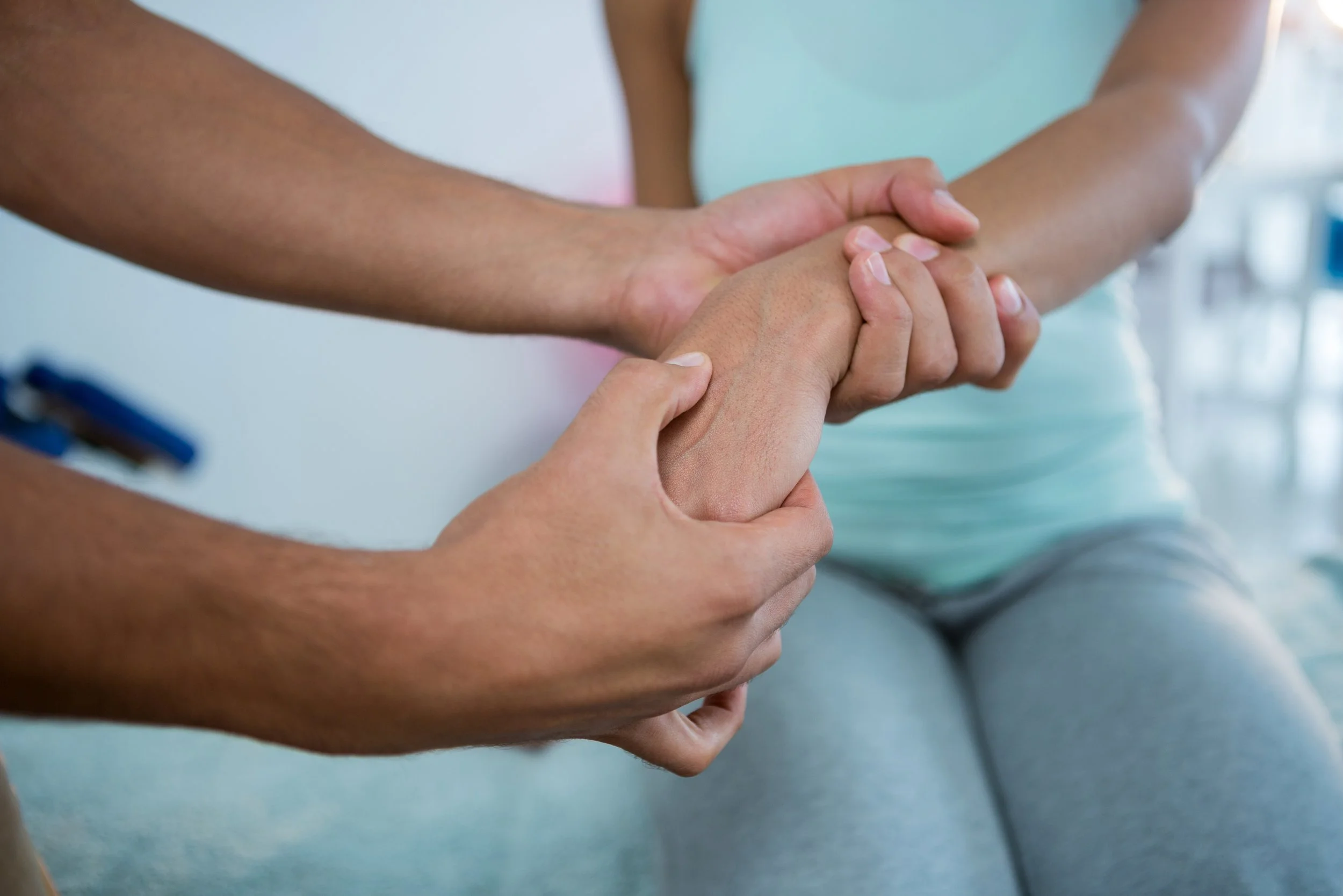
To diagnose your condition, your doctor will review your symptoms and conduct a physical examination.

To help diagnose the extent and severity of a brachial plexus injury, you may have one or more of the following tests:

  • Electromyography (EMG). During an EMG, your doctor inserts a needle electrode through your skin into various muscles. The test evaluates the electrical activity of your muscles when they contract and when they're at rest. You may feel a little pain when the electrodes are inserted, but most people can complete the test without much discomfort.
  • Nerve conduction studies. These tests are usually performed as part of the EMG, and measure the speed of conduction in your nerve when a small current passes through the nerve. This provides information about how well the nerve is functioning.
  • Magnetic resonance imaging (MRI). This test uses powerful magnets and radio waves to produce detailed views of your body in multiple planes. It often can show the extent of the damage caused by a brachial plexus injury and can help assess the status of arteries that are important for the limb or for reconstruction of it. New methods of high-resolution MRI, known as magnetic resonance neurography, may be used.
  • Computerized tomography (CT) myelography. Computerized tomography uses a series of X-rays to obtain cross-sectional images of your body. CT myelography adds a contrast material, injected during a spinal tap, to produce a detailed picture of your spinal cord and nerve roots during a CT scan. This test is sometimes performed when MRIs don't provide adequate information.
  • Angiogram. If your doctor suspects that the blood vessels feeding your arm might be injured, he or she might suggest an angiogram — an imaging test where contrast material is injected into an artery or vein to check the condition of your blood vessels. This information is important in planning your surgical procedure.

    This article originally appeared on mayoclinic.org

Nerve Pain: Pronator Teres Syndrome

Upper extremity nerve entrapments are a common cause of pain and disability.

The increase in repetitive motions associated with occupational and recreational environments usually is singled out as the primary cause of these problems.

Many individuals with nerve entrapment symptoms will seek the care of a massage practitioner.

pronatorteres.jpg

Figure 1. Anterior view of the left elbow showing the median nerve going under the superficial head of pronator teres. (3-D anatomy images courtesy of Primal Pictures Ltd., www.primalpictures.com.)

If a client comes to you with an upper extremity pain condition, you want to accurately identify that problem so you can determine if it warrants massage treatment or referral to another health professional. In some cases, a condition might have symptoms that very closely mimic a different pathology. If you don't identify the condition correctly, your treatment is not going to be as effective.

The symptoms of pronator teres syndrome (PTS) can be identical to those of carpal tunnel syndrome because they both involve compression of the median nerve. PTS may be underdiagnosed by medical professionals because its symptoms are so closely related to carpal tunnel syndrome, which is a much more well-known condition.1

PTS develops from compression of the median nerve by the pronator teres muscle, and is sometimes referred to as pronator syndrome. The term pronator syndrome also can include median nerve compression by other structures in the elbow, such as the ligament of Struthers or the bicipital aponeurosis (lacertus fibrosus).2

Figure 2. The sensory distribution of the median nerve in the hand.(Mediclip image copyright 1998, Williams & Wilkins. All rights reserved).

As the median nerve passes the elbow, it runs between the two heads of the pronator teres muscle, where the nerve may be compressed (Figure 1). Compression can be due to muscle hypertonicity or fibrous bands within the muscle pressing on the nerve.3 In some cases, pressure is placed on the nerve by anatomical anomalies, such as the nerve traveling deep to both heads of the pronator teres.4 In this situation, the nerve might be compressed against the ulna by the pronator teres muscle itself.

PTS results from repetitive motions that cause hypertonicity in the pronator teres. Occupational activities such as hammering, cleaning fish, or performing any activity that requires continual manipulation of tools can cause overuse of the pronator teres. The hypertonicity then causes nerve compression, and the symptoms are felt in the anterior forearm and the median nerve distribution in the hand (Figure 2). Women are affected more than men, although the reason for this is not clear.

Most symptoms of nerve compression radiate distal to the site of compression. Aching forearm pain and paresthesia, along with pain in the median nerve distribution in the hand, are likely to be PTS and should not be assumed to indicate carpal tunnel syndrome.

Figure 3. The pronator teres test.While PTS and carpal tunnel syndrome both affect the median nerve and have similar symptoms, there are distinct differences. PTS pain is exacerbated by repetitive elbow flexion, and symptoms arise in the forearm as well as the hand. Carpal tunnel syndrome is aggravated by wrist movements, and pain is not experienced as much in the forearm. In both cases, atrophy is possible in the thenar muscles of the hand, which are innervated by branches from the median nerve.

There are several other ways to identify PTS and distinguish it from carpal tunnel syndrome. Clients with carpal tunnel syndrome frequently report night pain, while individuals with PTS generally do not.1 Prolonged wrist flexion during sleep aggravates carpal tunnel syndrome because it decreases the space in the carpal tunnel and presses on the median nerve. Because wrist flexion does not affect the pronator teres muscle, this wrist position does not increase nerve compression symptoms in PTS.

An evaluation procedure called the pronator teres test also is helpful in identifying the condition. The client stands with the elbow in 90 degrees of flexion. The practitioner then places one hand on the client's elbow for stabilization and the other hand grasps the client's hand in a handshake position. The client holds this position as the practitioner attempts to supinate the client's forearm (forcing the client to contract the pronator muscles). While holding the resistance against pronation, the practitioner extends the client's elbow (Figure 3). If the client's pain or discomfort is reproduced, there is a good chance of median nerve compression by the pronator teres. The client should keep the elbow relaxed during the test, because holding the elbow firmly in flexion will not allow elbow extension.

Pronator teres syndrome is most commonly caused by muscular compression of the median nerve. Therefore, it is a condition that is effectively treated with massage. However, it is important that the practitioner accurately identify the problem so treatment can be directed to the proper region of the upper extremity.

This article originally appeared on massagetoday.com and was written by Whitney Lowe, LMT

What does it mean when Lymph Nodes are swollen? Can Massage Therapy Help?

Swollen lymph nodes facts

  • Lymph nodes, also referred to as lymph glands, are important part of the immune system.
  • Lymph nodes are located throughout the body, but visible and palpable only when they are enlarged or swollen.
  • Lymph nodes are regional, and each group of them corresponds to a particular region of the body and reflects abnormalities in that region. Common areas where swollen lymph nodes are more prominent and therefore more readily noticeable are behind the ear, in the neck, the groin, under the chin and in the armpits.
  • These are also usually the areas your doctor will check for lymph node enlargement.
  • In general, infections are the most common causes of lymph node enlargement. Other causes include inflammation and cancers. There is a wide variety of infections from a strep throat or ear infection, to mononucleosis or HIV infection, which can cause swelling of lymph nodes.
  • Lymphoma and leukemia cause swelling of lymph nodes, and many cancers spread to lymph nodes.
  • Rarely, a medication can cause swelling of a lymph node.
  • Symptoms associated with lymph node swelling and related diseases can include pain in the area of the swelling, fever and fatigue.
  • Not all swollen lymph nodes are abnormal.  

Want to learn more? Continue Reading


Once you've been checked out by a physician, ask them about receiving Lympathic Massage.

The Benefits of Lymphatic Massage

Discover How To Boost Energy and Immunity

Six months after hip replacement surgery, Larry was learning to walk again and life was returning to normal. But one thing still puzzled him. When he stood for any length of time, his left ankle would swell, and when the inflammation was at its worst, his right ankle would also swell.

"I can understand why my left leg is swollen," he says. "But why would my right leg swell? I didn't have surgery there. And why am I getting swelling six months after the surgery? Shouldn't it be better by now?" The answer is that although Larry's surgery had occurred on the opposite side, the right leg would swell when the inflammation became too much for the left side to handle.

Fortunately, lymphatic massage can help address Larry's problems. This special type of bodywork, while very gentle and seemingly superficial, helps to restore function to the lymph system and balance the body.

The Lymph System
Most people are familiar with the body's vessel system that carries blood to and from the tissues, but few understand there is another equally vital system of vessels that removes cell wastes, proteins, excess fluid, viruses, and bacteria. The lymph system picks up fluids and waste products from the spaces between the cells and then filters and cleans them.

Like the roots of a tree, the lymph system starts as tiny vessels--only a single-cell wide--that eventually branch into larger and larger tubes that carry these fluids back to the blood stream. This network of delicate vessels and lymph nodes is the primary structure of the immune system. The lymph nodes act as check points along the pathways of the vessels. They filter the fluid (called lymph) and serve as the home for lymphocytes--little Pac Man-like cells that attack and destroy foreign bacteria and viruses and even abnormal cells, like cancer cells.

When the lymph system works well, we feel healthy and have a strong defense against illness. When it's sluggish or blocked--say after surgery or an injury--we can have swelling, feel tired, and be more susceptible to colds and infections.

Lymphatic Massage
A customized form of bodywork, lymphatic massage may help the lymph system do its job better. By understanding the anatomy and function of this delicate system, your massage therapist can assist your body in clearing sluggish tissues of waste and swelling. 

Though lymph vessels are found throughout the body, most of them--about 70 percent--are located just below the skin. These fragile vessels work to pick up fluids between the cell spaces when gentle pressure is applied to them from increased fluid build-up, muscle contractions, or the pressure of a therapist's hands. By using very light pressures in a rhythmic, circular motion, a massage therapist can stimulate the lymph system to work more efficiently and help it move the lymph fluids back to the heart.

Furthermore, by freeing vessel pathways, lymphatic massage can help retrain the lymph system to work better for more long-term health benefits.

Massage therapists versed in lymphatic drainage therapy, an advanced form of lymphatic massage, can identify the rhythm, direction, and quality of the lymphatic flow and remap drainage pathways. 

Who Should Get It?
Lymph massage can benefit just about everyone. If you're feeling tired and low on energy, or if you've been sick and feeling like your body is fighting to get back on track, lymph massage would likely serve you well. 

In addition, athletes, surgical patients, fibromyaliga and chronic fatigue sufferers, as well as those wanting a fresh look may want to consider lymphatic massage. Here's why.

After a sports injury or surgery, lymph vessels can become overwhelmed with the demand placed on them. When tissues are swollen, deep tissue techniques may actually cause damage to the lymph vessels and surrounding structures. Lymphatic massage is often the treatment of choice, because it helps the body remove proteins and waste products from the affected area and reduce the swelling. This helps reduce pressure on cells and allows them to reproduce faster to heal the body.

Surgical procedures involving lymph node removal--such as breast cancer surgery--can cause limbs to swell. Severe limb swelling needs the attention of a medical team, but in milder cases, lymphatic massage alone may be enough to prevent or even treat the swelling. It's important that your doctor be involved in your care. Let your doctor know you'd like to see a massage therapist and make sure you have medical approval. 

Lymph massage can also be part of a care program for fibromyalgia or chronic fatigue syndrome. Because it's so gentle, it is well tolerated by these patients, who are often experiencing sore trigger points throughout the body. And by encouraging lymph flow and removing waste products, this gentle form of bodywork can help restore immune function and improve vitality.

Estheticians are trained in a very specific form of lymphatic massage. When you get a facial, your esthetician will gently massage your face to help improve lymph flow. When lymph is moving freely in the face, you'll have clearer, healthier skin without a buildup of toxins and fluids.

So, if you're feeling a bit sluggish, experiencing mild to moderate swelling, recovering from a sports injury, or interested in optimizing your lymph system for stronger immunity, ask your massage therapist about lymphatic massage. It can have a powerful impact on your body's ability to heal. 

This article originally appeared on massagetherapy.com and was written by Cathy Ulrich.


If We Can't Stretch Fascia, What Are We Doing?

When Ida Rolf (developer of the profound therapy, Rolfing) began putting her hands and elbows on people’s skin and applying pressure, creating a slow, sustained stretch, she imagined that she was stretching fascial sheets. Generations of manual therapists have followed her thinking, accepting this explanation to account for the changes felt in tissue tension beneath their hands and the sensations experienced by those who receive this type of therapy.

Ideas change over time

Much of manual therapy has grown largely out of anecdotal experience and tradition. Without the means to directly observe or measure what happened inside of the body, explanations for results had to be created from the “outside” and have largely been guesswork. As manual therapy has moved forward, an interest in understanding exactly how touch affects the body has led to a growing interest in research. With research has come the realization that many explanations of the past are not supported by evidence and are sometimes contradicted by evidence. Science-minded manual therapists have learned to adapt to this information, dropping outdated hypotheses and unsupported claims. While some have found it disconcerting to have cherished notions disproved, others have embraced knowledge and have adapted their conceptual models to fit what is known. They may continue to use modalities that have produced desired results but their understanding of how that comes about changes to fit the evidence.

Such a change is happening in the field of “fascial” therapy. 

When Rolf began her groundbreaking work in manual therapy, she devised a hypothesis in an attempt to explain how changes created by her contact came about. However, in recent years, evidence has challenged those explanations. Robert Schleip, Ph.D., was one of the key organizers of the first Fascia Research Congress and is a highly respected researcher. He is credited with discovering minute contractile fibers in fascia, a discovery whose clinical relevance has not yet been demonstrated but still excited many in the world of fascial therapy just the same. In his two-part article, “Fascial Plasticity: a new neurobiological explanation,” published in 2003 in the Journal of Bodywork and Movement Therapies, Schleip points to studies which contradict the notion that we can change the shape of fascia with our hands. One study found that collagen fibers would only begin to stretch shortly before they reached the breaking point, something that would not be desirable in a living human being. In other studies, Schleip, Trager, and others have done Rolfing under anesthesia and found that it produced no results. If the application of manual pressure had the ability to stretch fascia, there should have been a change in spite of anesthesia blocking any neural response. Why, then, was there no change when anesthesia took the nervous system out of the picture?

A neurobiological explanation

If we aren’t stretching fascia, then how do we account for the “release” felt by both the practitioner and the subject? Schleip and others have suggested that the change in tonus is not achieved by an alteration in the shape of fascia but is instead controlled by the nervous system. Schleip suggests that one possible mechanism of change brought about by sustained manual pressure could be the Ruffini corpuscles.

Why Ruffini corpuscles? Clinically, we observe that applying a slow, extended stretch to the skin can create desirable changes both locally and centrally, decreasing tension in the area where the hands are applied as well as creating an overall sense of relaxation. Ruffini corpuscles respond to lateral skin stretch, that is, stretching the skin tangentially or along the same plane as the tissue below. They are slow-adapting, meaning that they continue firing for as long as the stretch is sustained, unlike some mechanoreceptors which respond briefly to new stimulation and then stop responding if it continues.

We know that when we apply our hands to the skin of the body, we stimulate mechanoreceptors. Impulses are sent through the sensory nerves to the brain. The brain evaluates and responds, sending out impulses of its own through nerves to various parts of the body, causing changes to occur in the diameter of blood vessels, breathing, muscle tonus. If it likes our touch, it can create the changes we associate with relaxation, release of tension, and can decrease the sensation of pain. If it feels threatened by our touch, it will do the opposite. As manual therapists, we are always trying to create changes that make the body feel at ease. We can achieve this through the nervous system.

The nervous system is constantly monitoring its environment, responding to a complex array of input. It would be naive and simplistic to think that response to our touch could be reduced to one set of mechanoreceptors or to ignore all the other countless factors. However, when examining the kind of manual therapy we have come to think of as "fascial," understanding the role of Ruffini corpuscles is a good place to start.

Why does it matter?

Does it matter whether we believe we are stretching fascia or not? It matters that we think accurate thoughts about how the body works and what effect our touch has on the body. Understanding how the body actually works will help us work more effectively.

We may still use our hands in ways that we have before. If those methods work to achieve the client's goal, there is no need to abandon them. However, we want to know that how we think about what we are doing is accurate and we want to be able to communicate honestly with our clients. If we discover that our conceptual model is contradicted by what is known about how the body works, then it is time to adapt our model so that our thinking is in agreement with evidence.

Manual therapists need not feel threatened by the news that we cannot stretch fascia. A growing number of Rolfers, practitioners of myofascial release, and related modalities are continuing to use their hands in the ways that have worked for them in the past while adapting their thinking to an updated neurobiological explanation. Many have found that this shift to thinking about the role of the nervous system in manual therapy has led to new, even more effective approaches.
 

A thought experiment

Schleip proposes an interesting thought experiment. During the time it took to read this article, one’s bottom, if seated, is subjected to more pressure over a longer period of time than most therapists will apply to the hips of a client. Yet most of us are not all stretched out and droopy from daily sitting for extended periods of time. Think about it.

This article originally appeared on www.massage-stlouis.com and was written by Alice Sanvito, LMT